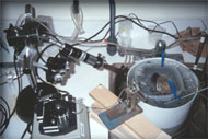
Video endoscopy has been used in the medical and veterinarian fields for decades to examine internal organs and structures of humans and animals. Its use in marine biology, however, was limited due to cost, lack of techniques, and methodological problems. In 1990, while employed as a post-doctoral Research Fellow at Memorial University, Newfoundland, Canada, my colleagues and I developed techniques to apply video endoscopy to the study of bivalve molluscs. Since that time, the observations and data we have collected from in vivo observations of feeding have shed new light and understanding on the process of particle feeding in bivalves and other suspension feeders.
Numerous conference presentations, invited seminars, and publications have resulted from this work (see Publications). In addition, we have developed an educational video that demonstrates feeding processes in bivalves, and I have participated in the making of an Open University, teaching video that explores the functional biology of suspension feeders.
Note: All video clips are protected by international copyright laws and are intended for use by individual researchers or instructors in a class-room setting. Those wishing to use the video clips for other purposes should contact me for permission.
A. Videos for general interest and education
B. Video produced by the BBC for the Open University (UK)
C. Videos to support data and interpretations presented in Ward et al. (1998, L&O, 43: 741-752) and Ward et al. (2000, L&O, 45: 1203-1210)
These video sequences demonstrate the important points concerning in vivo observations of feeding and our particle capture model. Please see the above papers for a full explanation of data presented in the video clips.
Note: Video clips have been compressed to reduce disk storage space and down-loading time. For best viewing, dim room lights and adjust monitor brightness and contrast if needed. Quality of images will vary depending on quality of the video card and monitor of your computer system. Original images are of higher quality.
- Quality 1: Demonstration of high quality images obtained with modern endoscope system.
- Quality 2: Demonstration of low quality images obtained with old endoscope system.
- Resolution 1: Calibration of endoscope system showing 3.0 um as limit of resolution.
- Resolution 2: Demonstration of some aspects of particle capture and visualization of movement of individual particles and phytoplankton cells.
- Dye Study: Demonstration of flow characteristics within the pallial cavity.
- Particle Tracking: Demonstration of particle kinematics above and along the ctenidia of bivalves. Stop-frame analysis allows us to track particle trajectories and movements.